Retinal Laser Photocoagulation
Ophthalmologists employ retinal laser photocoagulation to treat a wide range of retinal conditions.

Retinal Laser Photocoagulation
What is Retinal Laser Photocoagulation?
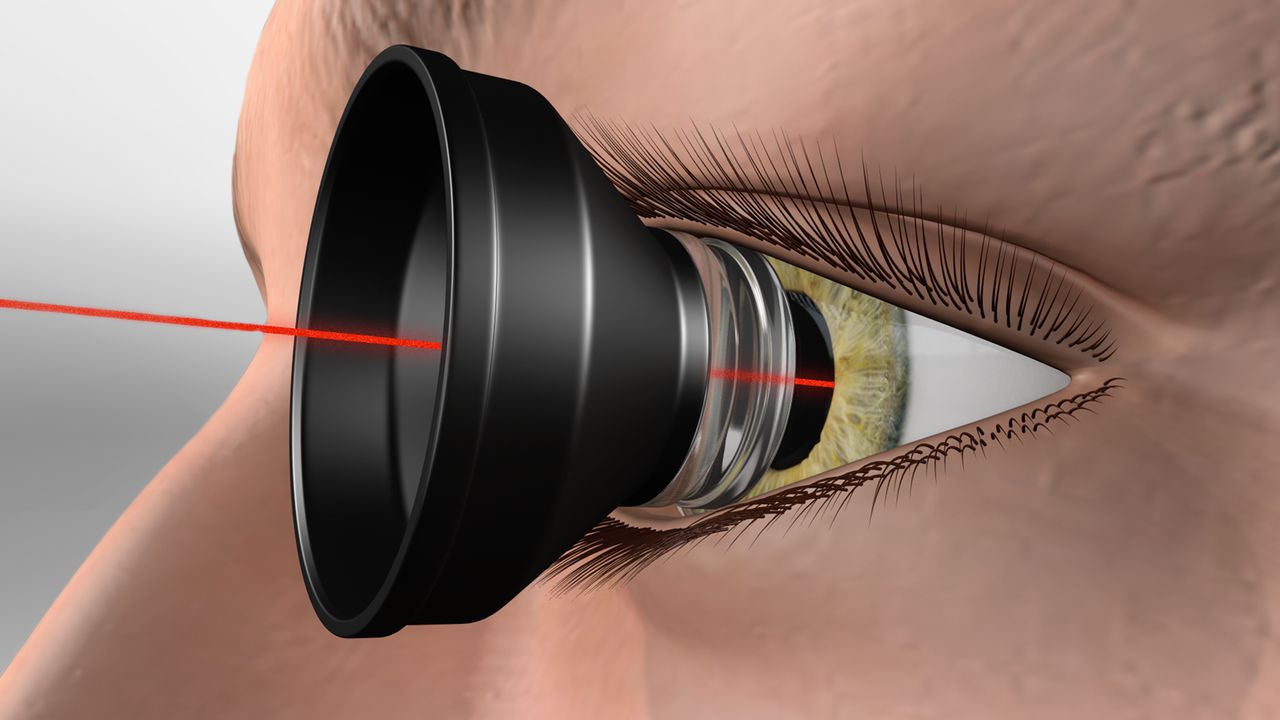
Ophthalmologists employ retinal laser photocoagulation to treat a wide range of retinal conditions. Diabetic retinopathy, retinal vein occlusion, retinal tears, central serous chorioretinopathy, and choroidal neovascularization are among the conditions on the list. The technique is not like surgery, contrary to the patients’ expectations. The doctor doing this treatment directs the laser beam (focused light waves) to the correct spot on the retina. The treatment is administered by creating heat energy, which causes the retina to coagulate.

Types and benefits of Retina Laser
According to the type of retinal disorder, laser therapy is provided in different ways.
Proliferative Diabetic Retinopathy (PDR)
Retinopathy caused by diabetes can progress to a more severe form called proliferative diabetic retinopathy, which is considered end-stage. Retinal blood vessels undergo alterations due to long-term diabetes and excessive blood sugar levels, ultimately resulting to PDR. Progressive retinopathy of the eye (PDR) is a serious condition that can cause blindness. Eye problems, such as bleeding from the aberrant arteries and/or retinal detachment, might arise if therapy is delayed.
When it comes to PDR, retinal laser therapy is effective because it lessens the likelihood of problems. Pan-retinal photocoagulation (PRP) is a procedure performed by the doctor to treat PDR.
Vision is the work of the retina, a structure that covers the whole back of the eye. The macula, located in the retina's centre, is the primary region responsible for high resolution vision. Doctors treating proliferative diabetic retinopathy avoid damaging the macula by focusing laser therapy on the retina's less-vascular regions. Since the almost 360 degrees of retina is slowly filled with laser spots, treatment for proliferative diabetic retinopathy can be completed in three to four sessions. This method helps avoid problems and irregular blood vessel development.
Diabetic Macular Edema (DME)
The macula swells and vision is impaired due to DME, an abnormal collection of fluid that can be caused by a variety of factors. Some cases of DME can benefit from retinal laser photocoagulation. In this case, the enlargement of the macula is reduced with the use of pinpoint laser treatments.
Retinal Vein Occlusion (RVO)
Retinal vascular occlusion (RVO) occurs when blood flow to the area of the retina fed by a particular vessel is disrupted for any number of causes. Retinal laser therapy is effective in this context, just as PRP is in PDR.
Retinal Tears, Holes and Lattice Degeneration
Nearly 10% of the general population, and especially myopes, suffer from retinal tears, holes, or lattice degenerations (areas of retinal thinning). Retinal detachment can occur through the tears if they are left untreated.
With two or three rows of laser spots around the breaks, the doctor can delimit the retinal breaks, resulting in thick adhesion in the surrounding retina and reducing the chance of retinal detachment. Screening for and laser removal of such lesions is required prior to LASIK and cataract procedures.
Central Serous Chorioretinopathy (CSC) and Choroidal Neovascularization
Both the conditions lead to areas of leak at the macular level, causing fluid collection and vision loss. Based on the specialist’s decision, in some cases, retinal laser therapy targeting the leaky areas is beneficial.
Patient preparation
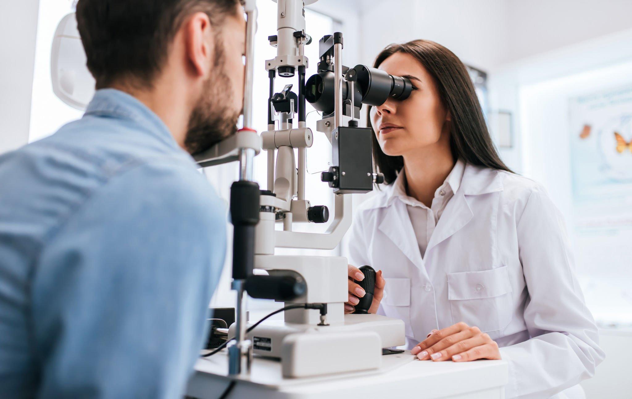
Only after topical anaesthesia has been applied can the laser process begin. To lessen discomfort, eye drops would be given beforehand. The process causes minimal discomfort. In some cases, the therapy may cause a slight prickling sensation for the patient. Depending on the severity of the patient’s illness, the entire process could last anywhere from five to twenty minutes.
After the procedure
For a day or two, the patient may experience some light glare and visual discomfort. Depending on the surgery, he or she may be instructed to use antibiotic and lubricant eye drops for anywhere from three to five days. A loss of contrast sensitivity and colour vision may result from severe PRP in diabetic retinopathy.
Types and method
Laser therapy can be administered in two distinct ways: through contact and without physical contact. A contact lens coated with a lubricating gel will be placed over the patient’s eyes during the contact process, and laser therapy will be administered while the patient is seated.
The patient is positioned prone before laser therapy is administered in this non-contact procedure. With a portable tool, the doctor may occasionally apply light pressure in and around the patient’s eyes.
Conclusion
To treat retinal tears, retinal laser photocoagulation is a quick, non-invasive, and painless option.
